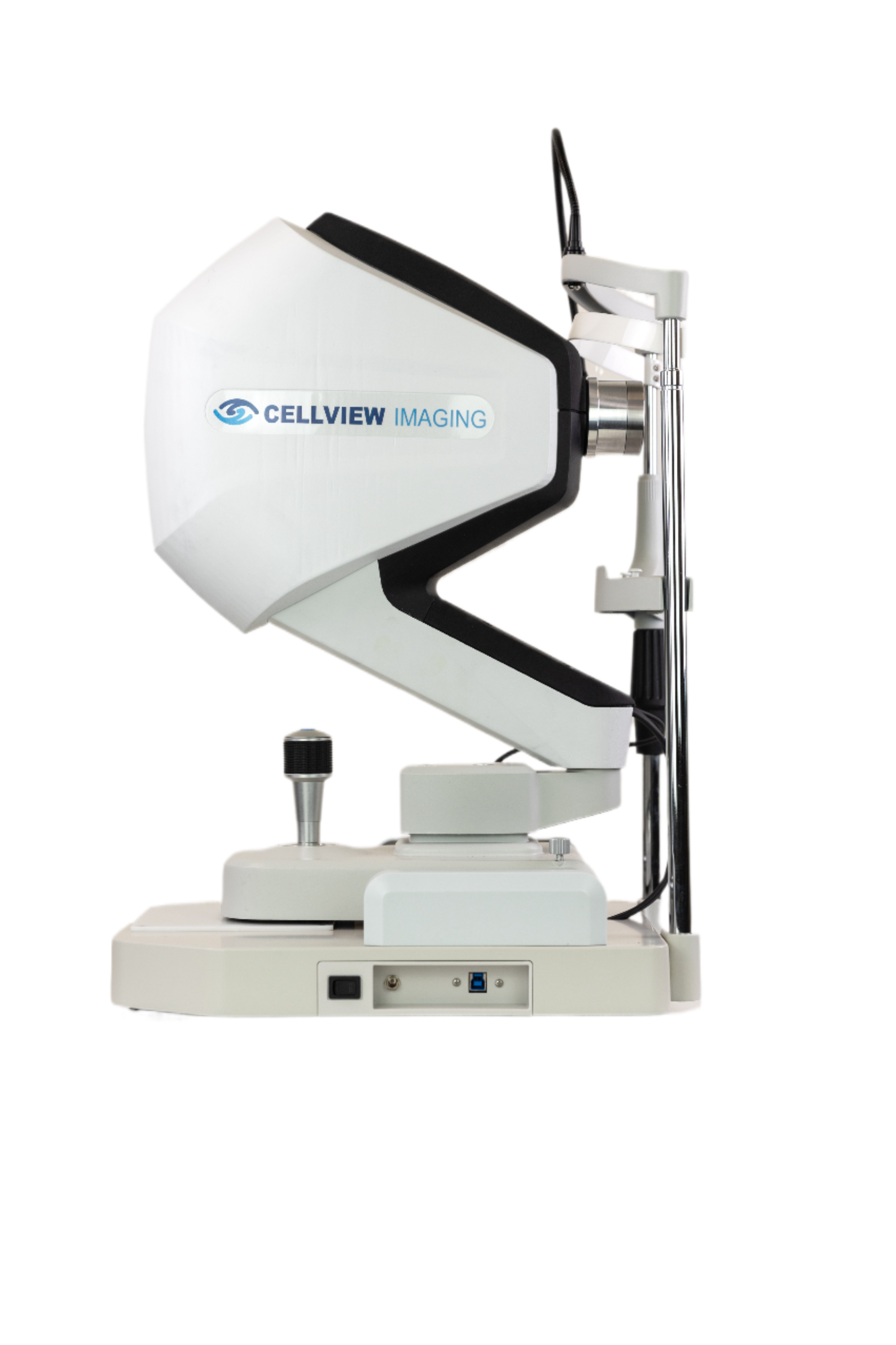
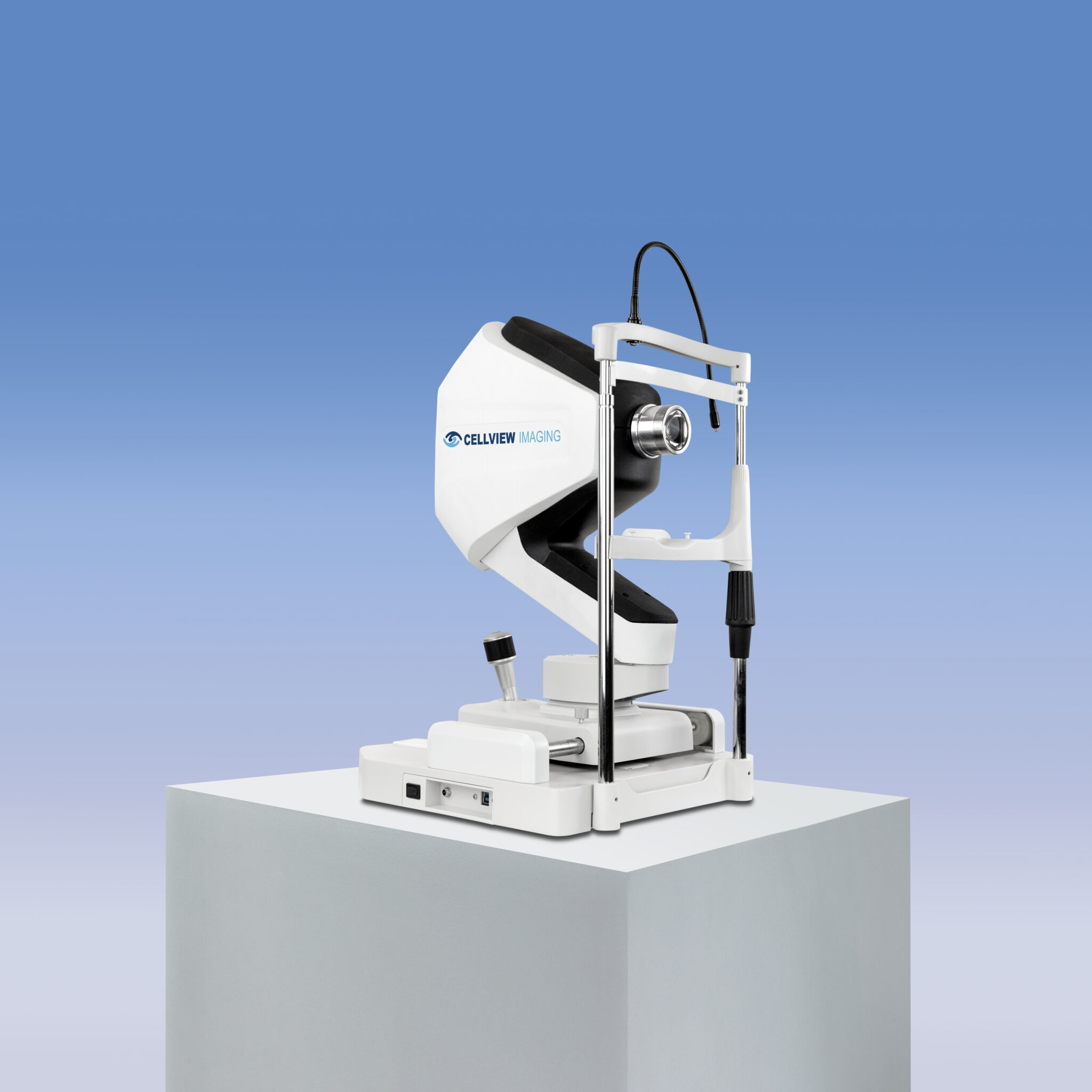
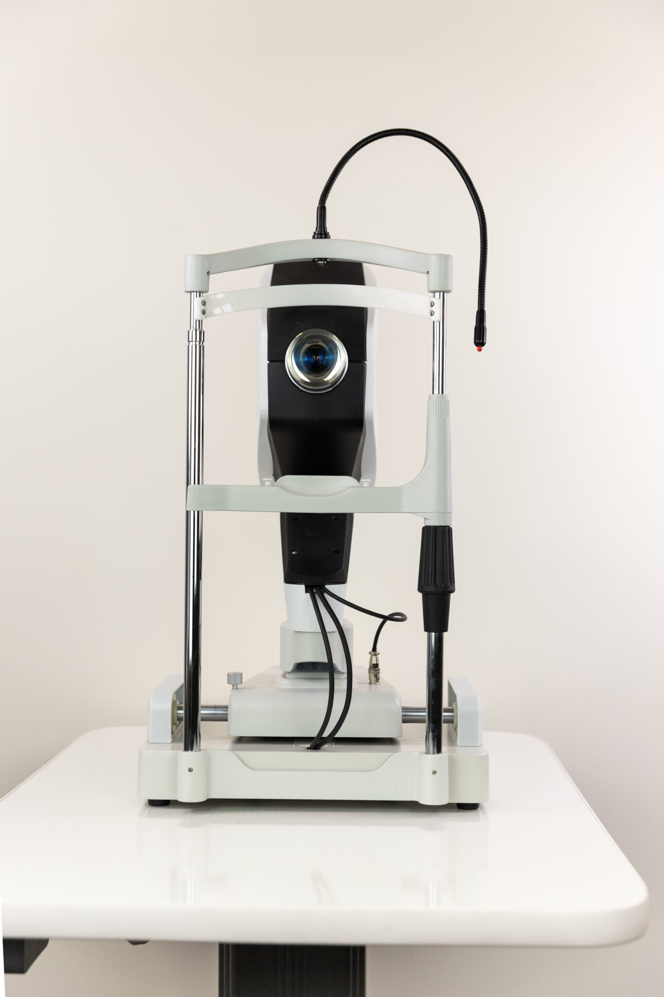
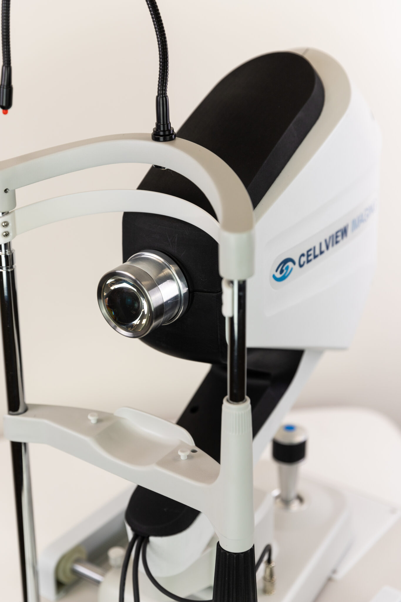
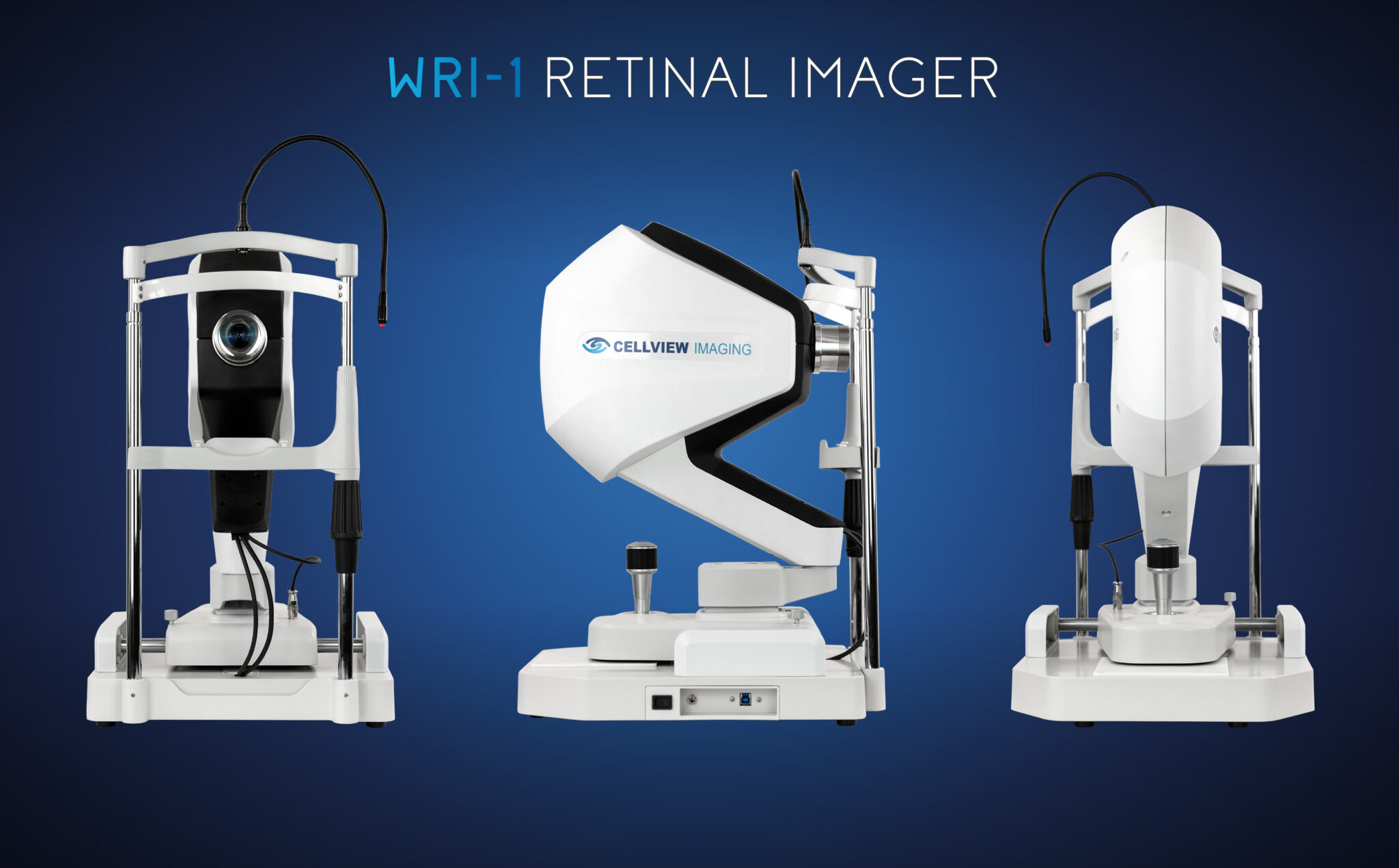
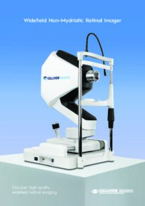
Widefield non-mydriatic retinal imager
Discover high-quality widefield retinal imaging
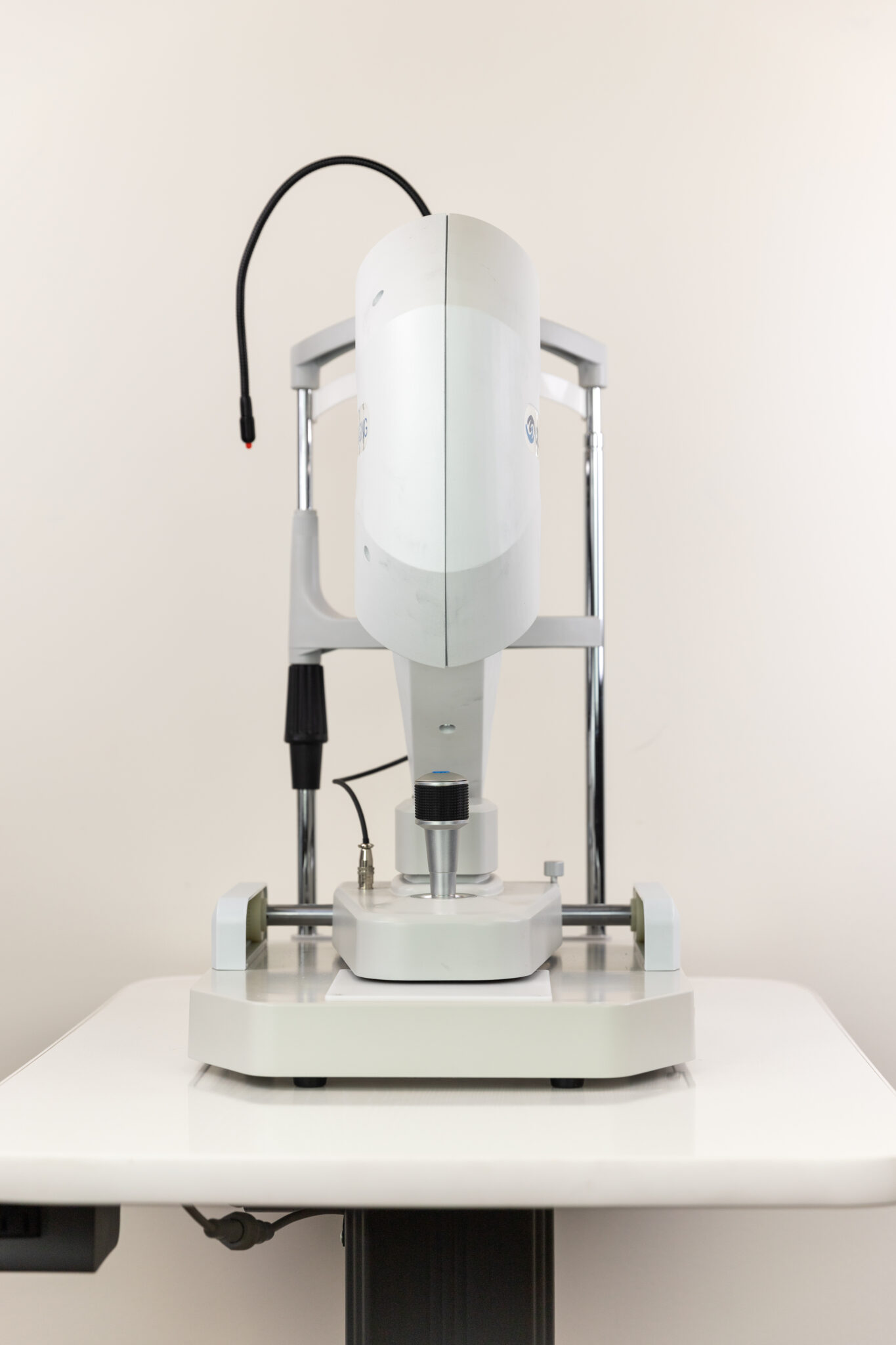
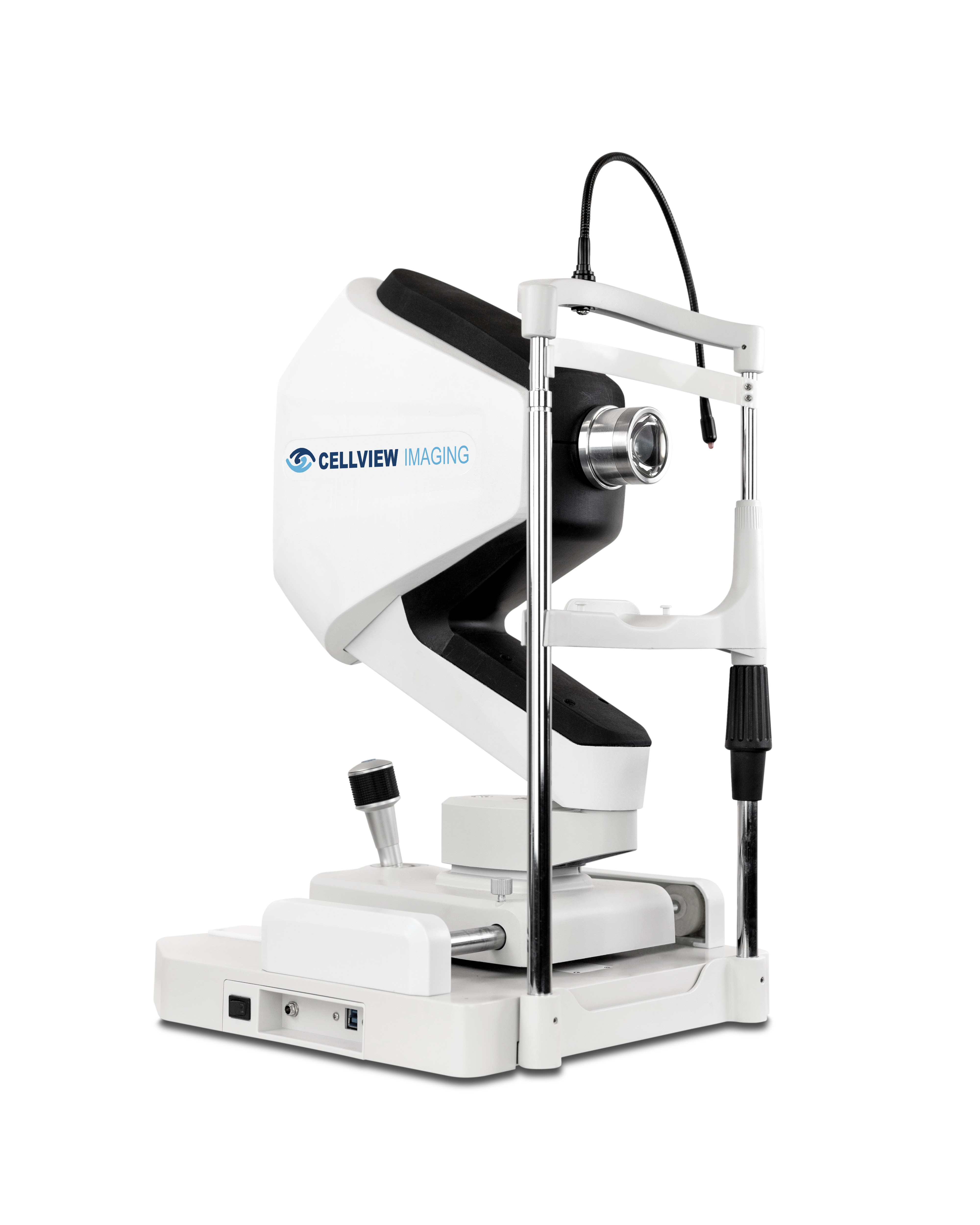
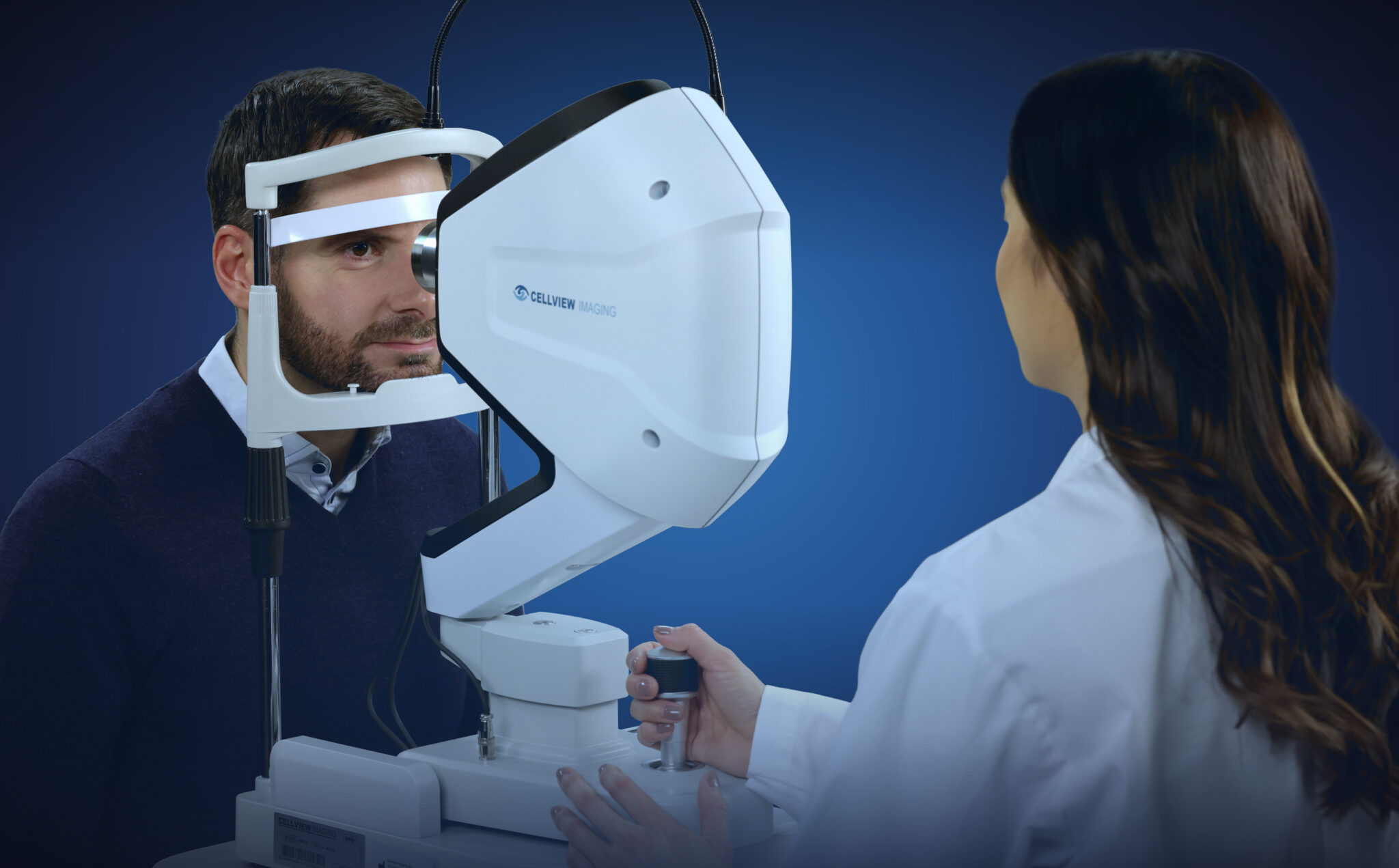
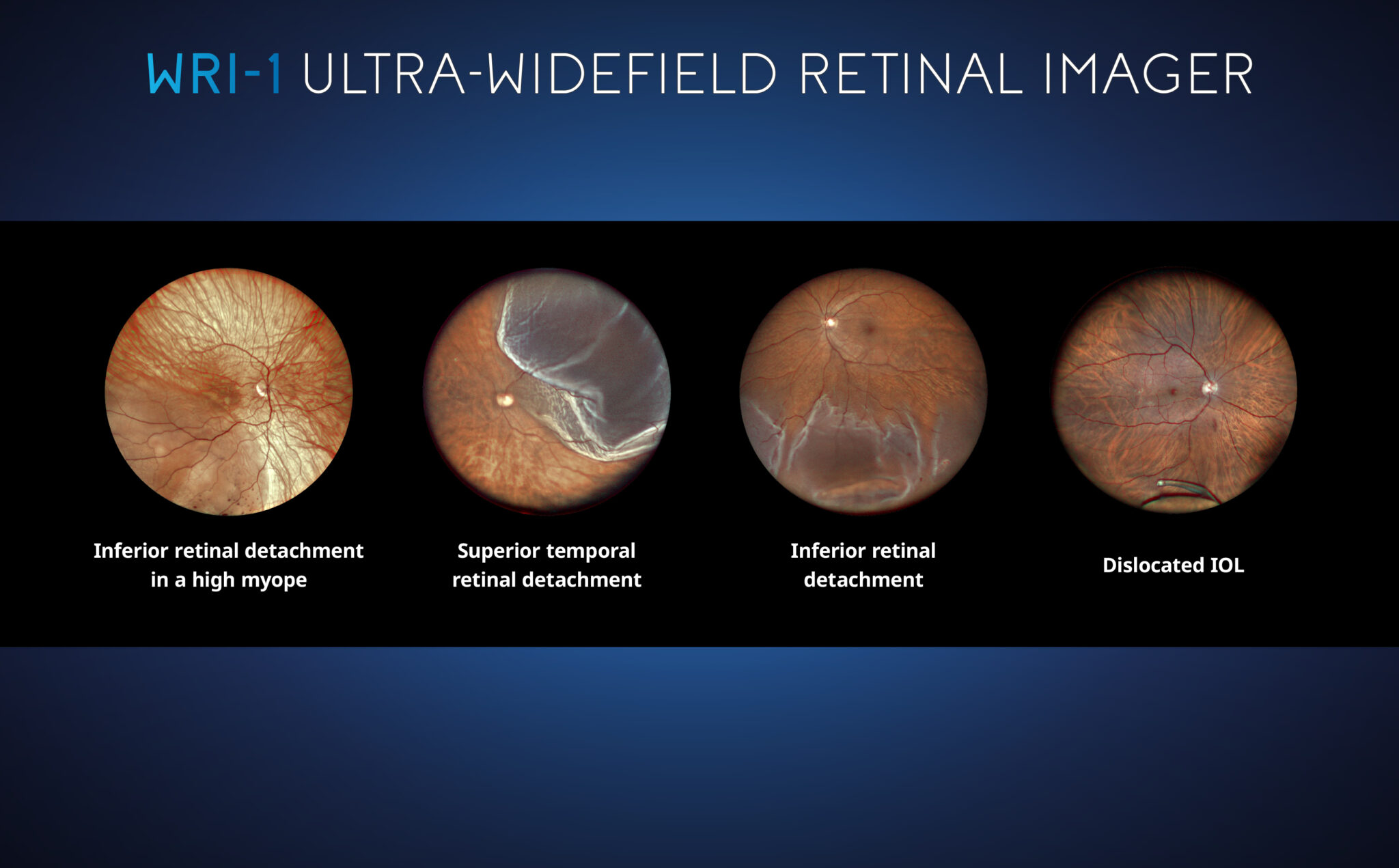
WRI-1 from Cellview captures a retinal image and far into the periphery, up to 133° in a single-capture, or an up to 200° auto-stitched image. It provides clear and high-definition images through small pupils, most cataracts, and other media opacities.
- Full visible spectrum LED array, with true color and infrared imaging, to allow for more accurate identification of potential pathologies that may go unnoticed with other imaging methods.
- Easy-to-operate for nominal staff training, unskilled operator usage, and decreased examination times for greater practice throughput.
![]()


Request PricingAdd to cart
Note: Submitting a support ticket will take you to a separate website.


Cellview WRI-1 Retinal Imager Brochure
Cellview Image Capture Guide
Cellview Practitioner’s Guide
We’ve been very impressed with the Cellview retinal camera, particularly its ease of setup and day-to-day use. Both staff and doctors can be trained quickly with minimal effort, which has made integration into our workflow seamless. I also perform portable eye exams, and the camera’s design makes it extremely convenient to transport without sacrificing functionality. On top of that, the cost point is excellent for the value it provides, making it a highly practical option for practices looking to expand imaging capabilities efficiently.
Download testimonial
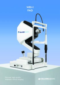
Cellview WRI-1 Retinal Imager – FAQ
Please Contact Us for the user manual for the Cellview WRI-1 retinal imager.