OCT500 Optical Coherence Tomographer
Optical coherence tomography (OCT) with integrated fundus imaging
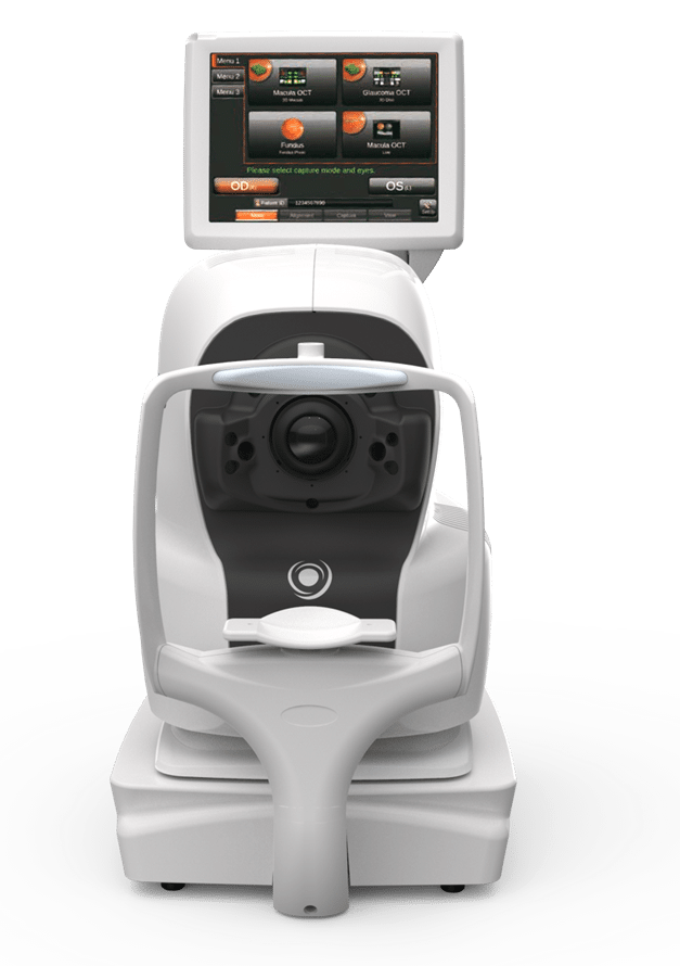
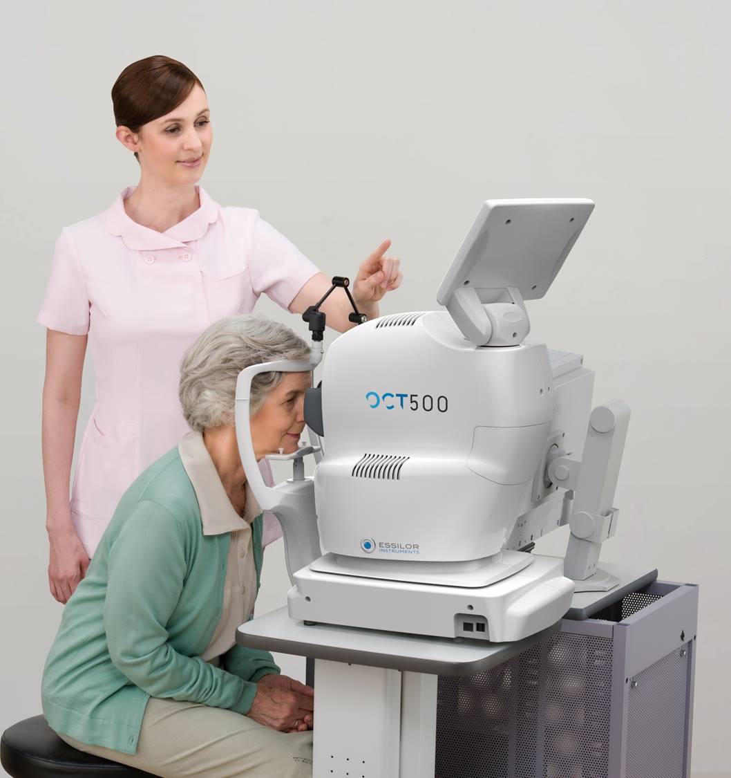
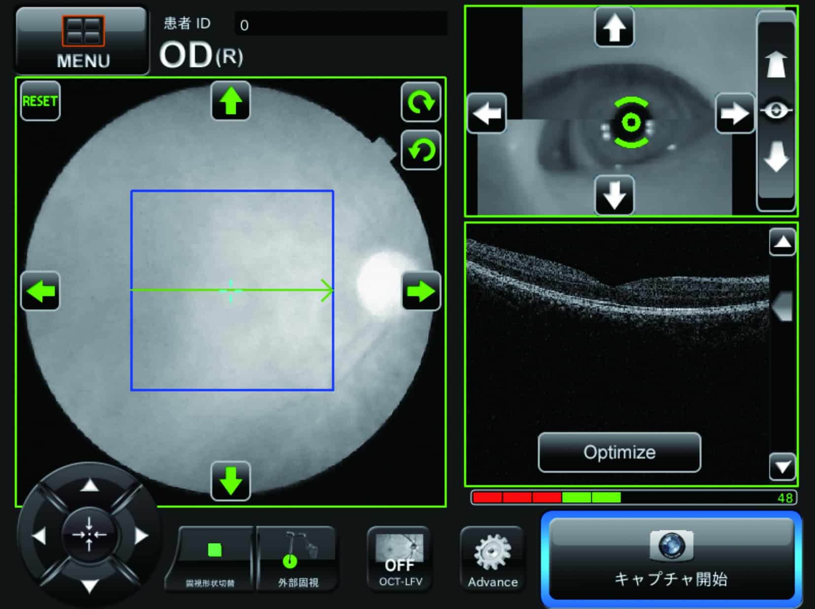
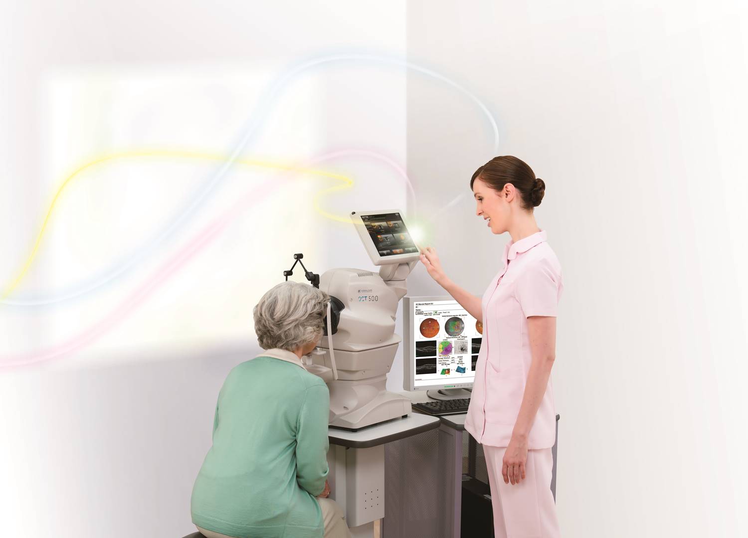
The OCT 500 optical coherence tomographer is the most user-friendly OCT on the market due to its fully-automatic function. With one touch on the screen, eye focus, optimization, and image capture are performed automatically. After capturing, the report can be displayed immediately by clicking on a single icon.
- Superb OCT technology ready for delegation
- Fully-automated operation with follow-up scan function
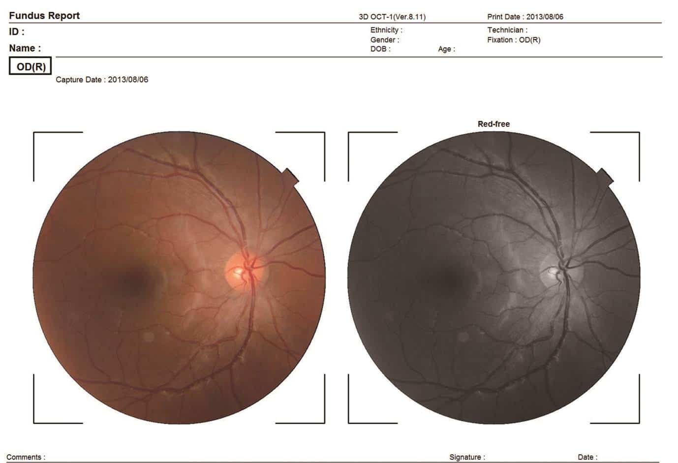
- True-color fundus photography
- Extended range of analysis function for the pupil and macula
- Compact and space-saving design
- Network and DICOM connectivity
Note: Submitting a support ticket will take you to a separate website.
")
Request PricingAdd to cart
Click for full view:
-
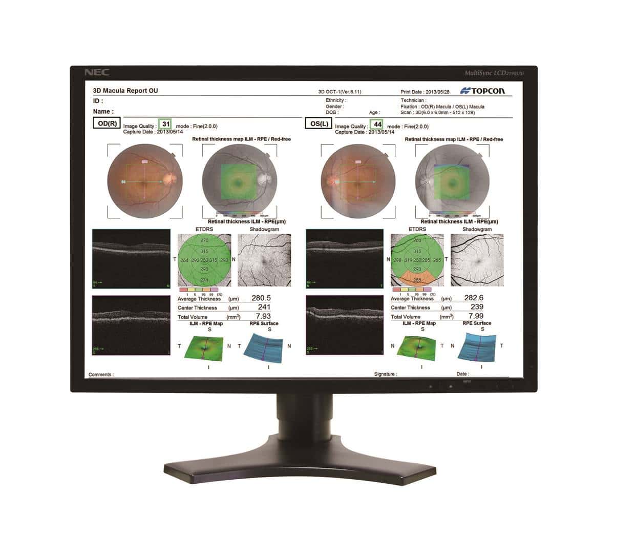
- Report Display
-
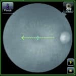
- Semi-Automatic or Manual Capture
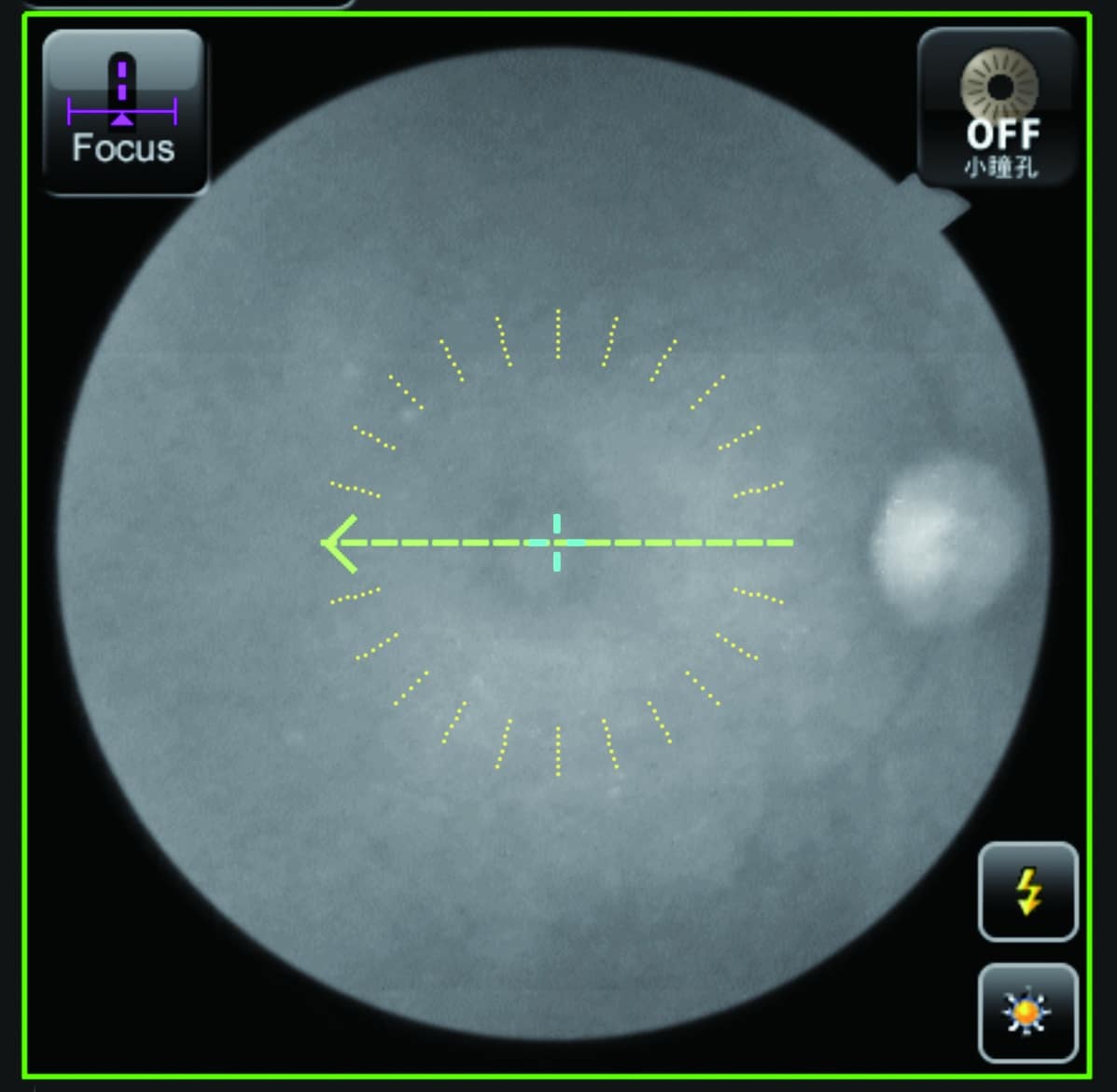
-
- Semi-Automatic or Manual Capture
-
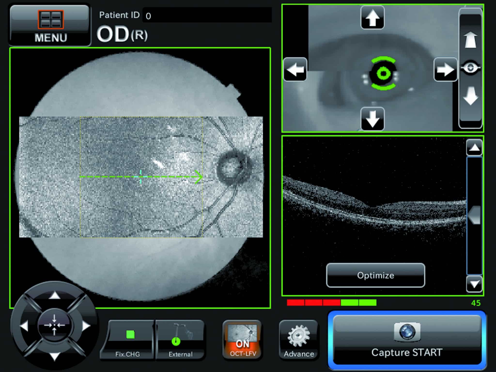
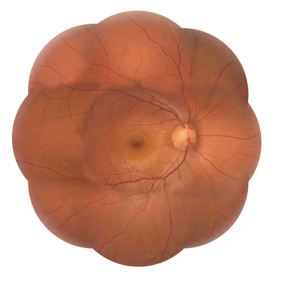
- Live Fundus View
-
- Live Fundus View
-
- Peripheral Fundus Photography
-
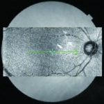
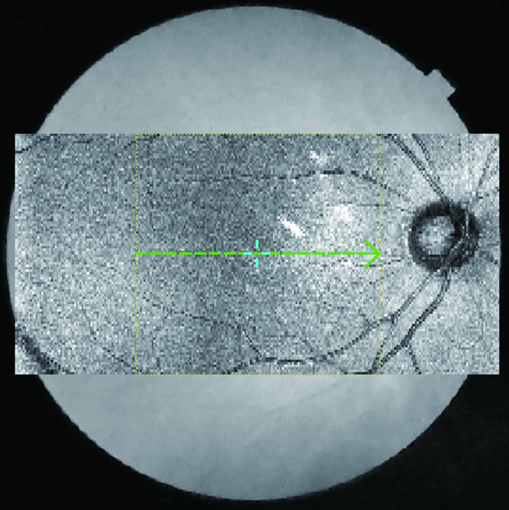
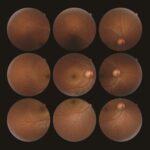
- Peripheral Fundus Photography (Mosaic)
-
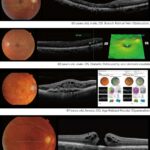
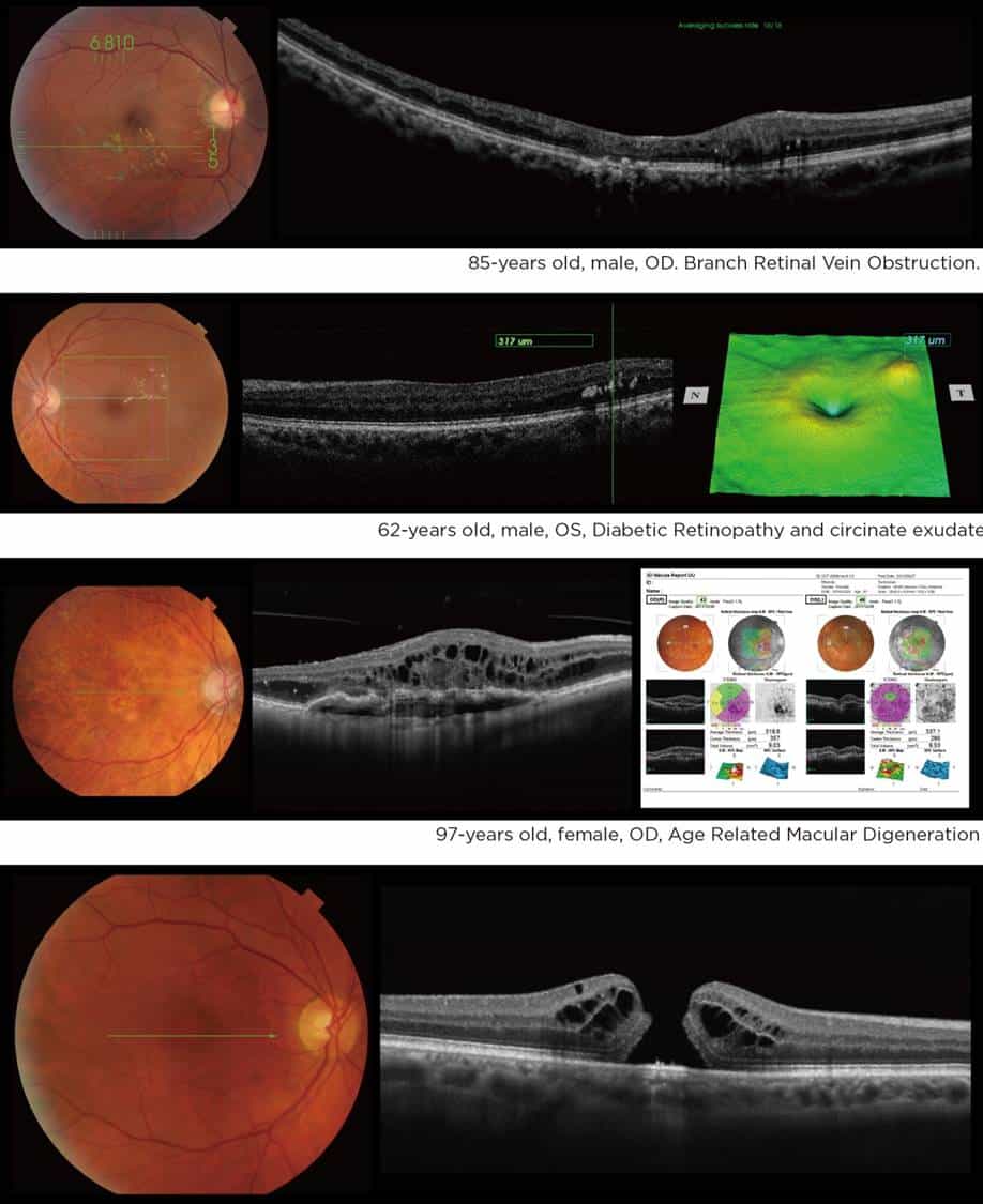
- Sample Patients
-
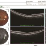
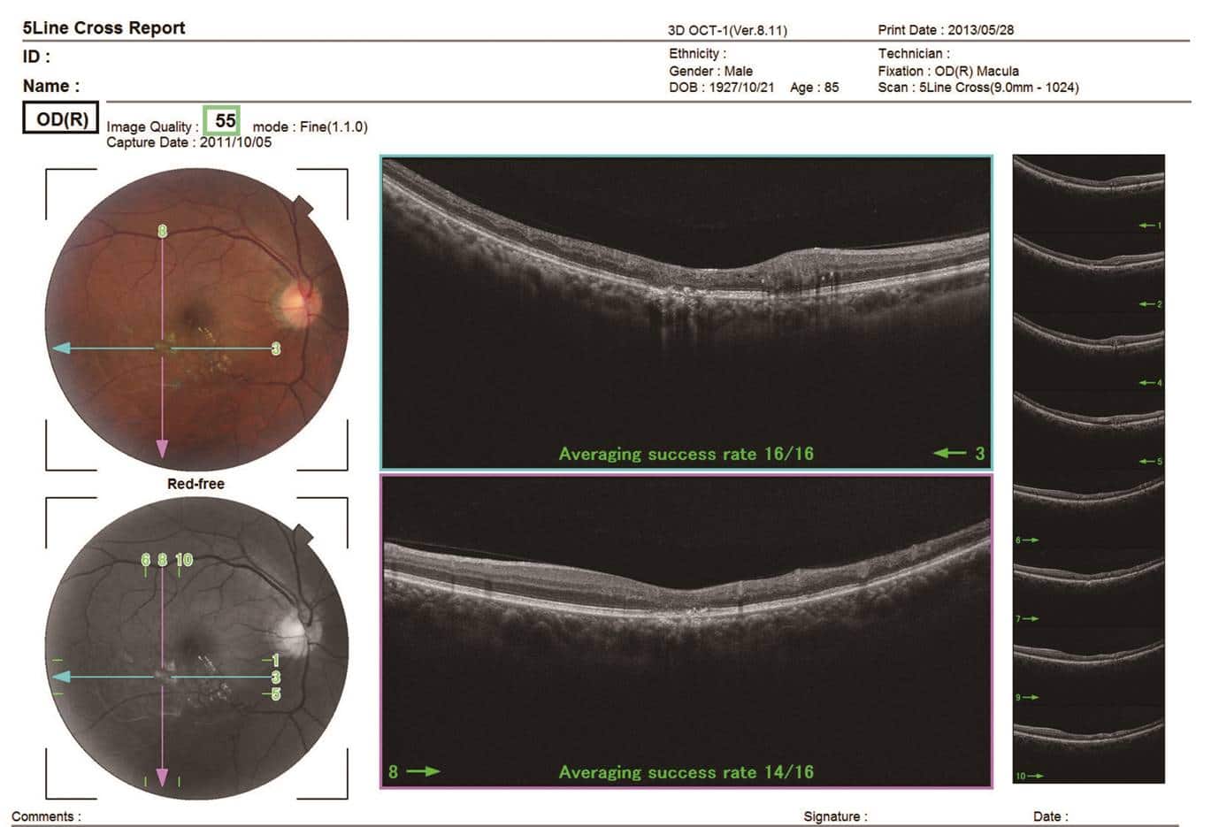
- 5 Line Cross Scan
-
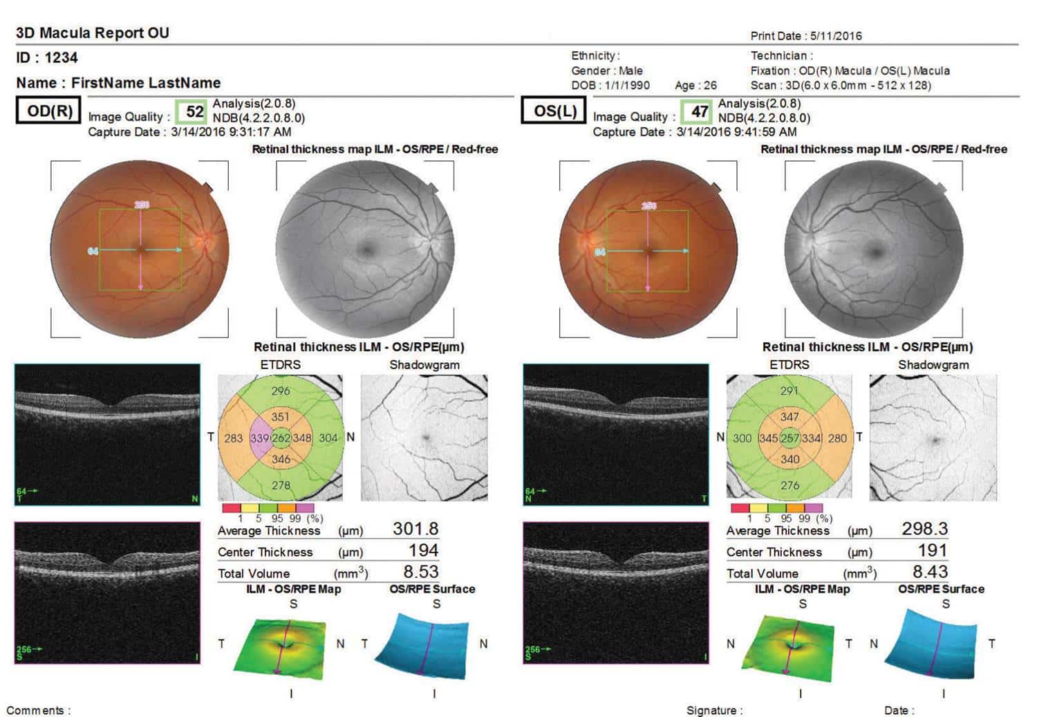
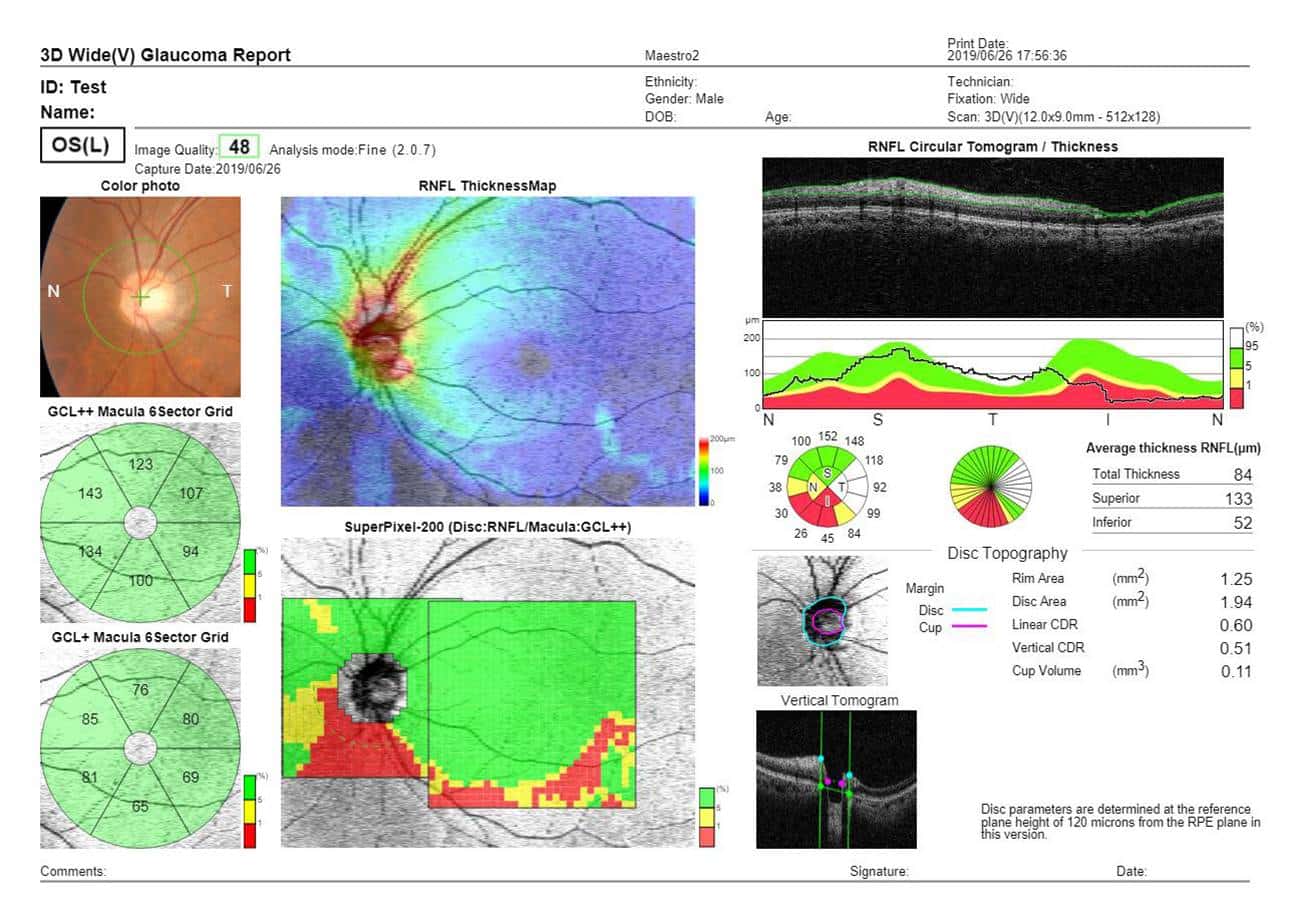
- 3D Macular Analysis
-
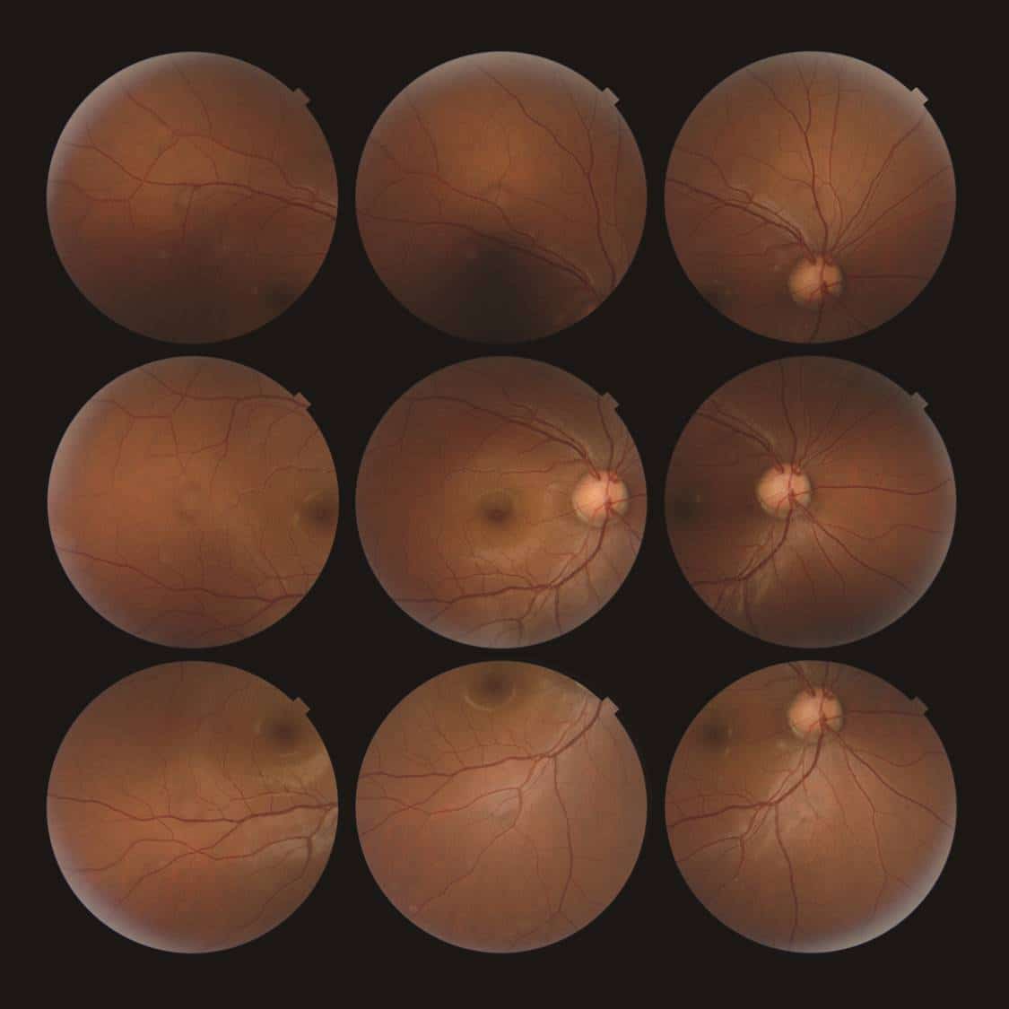
- Peripheral Fundus Photography (Right & Left)
-
- 3D Wide Scan (12mm x 9mm)
-
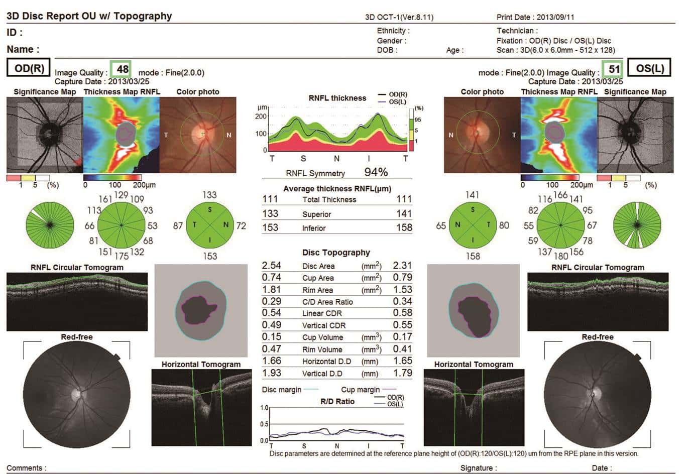
- 3D Disc Analysis
-
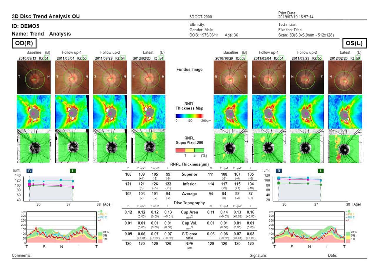
- Trend Analysis (RNFL)


OCT 500 Advanced Robotic OCT brochure